
Early Detection of Skin Cancer Through Noncontact Infrared Thermography
Identifying Subtle Heat Changes for Early Skin Cancer Diagnosis in Research with Infrared Microscopes
Thermal Signature of Skin Cancer: Exploring the Heat Variations in Cancerous Tissues
Skin cancer is a type of cancer that begins in the skin’s cells, often as a result of prolonged exposure to ultraviolet (UV) radiation from the sun or artificial sources like tanning beds. The disease develops when DNA in the skin cells is damaged, leading to abnormal cell growth. Skin cancer is primarily classified into three types: basal cell carcinoma (BCC), squamous cell carcinoma (SCC), and melanoma. BCC and SCC are more common and typically less aggressive, while melanoma is less common but significantly more dangerous due to its high potential to spread to other parts of the body. Skin cancer usually presents as changes in the skin, such as new growths, sores that do not heal, or alterations in existing moles. Early detection and treatment are vital, as many skin cancers can be successfully treated if caught early, especially the less aggressive forms.
Cancerous skin generates more heat due to several physiological changes linked to tumor growth. One of the main reasons is the increased metabolic activity of cancer cells. These cells have a higher metabolic rate than normal cells because they divide rapidly and require more energy, producing more heat as a byproduct.
Another factor is angiogenesis, the formation of new blood vessels, which tumors induce to supply the rapidly growing cancer cells with the necessary oxygen and nutrients. This increased blood flow to the tumor area often results in higher temperatures in the surrounding tissue. Additionally, inflammation commonly occurs around cancerous tissues, contributing to elevated local temperatures. The immune response to abnormal cancer cells triggers inflammation, leading to further heat production in the affected area.
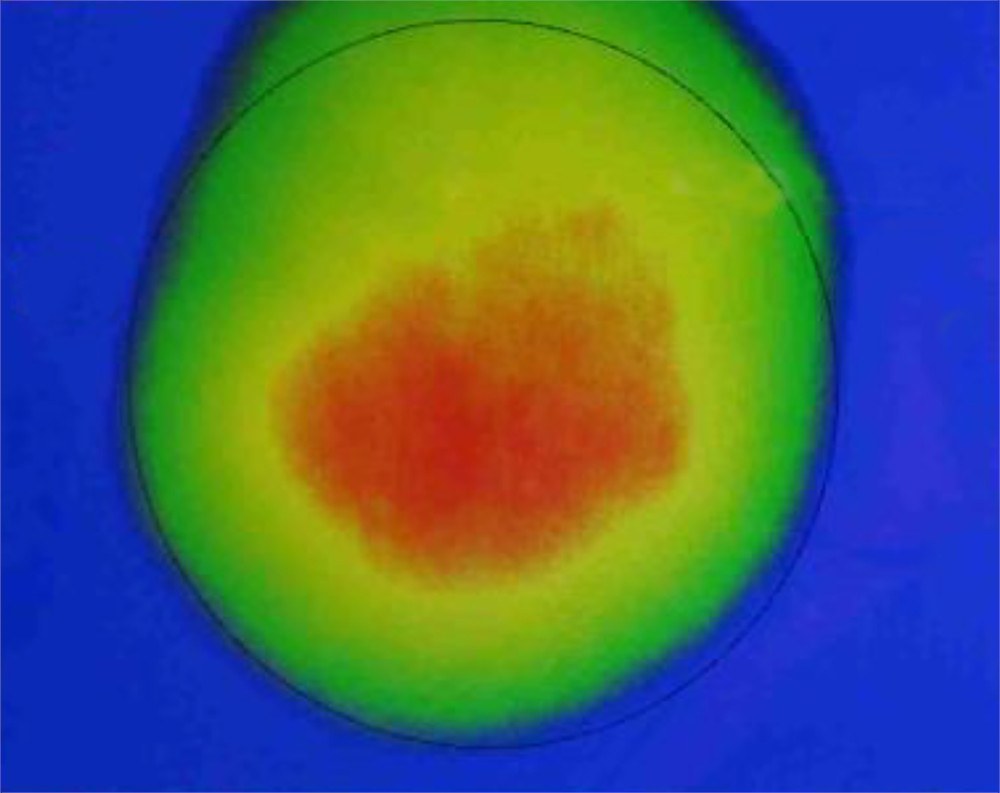
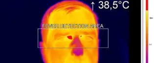
The temperature difference between cancerous and normal skin can vary, but it is typically about 1 to 3 °C higher in areas affected by cancer.

Infrared Thermography in Skin Cancer Research: Detecting Heat Signatures for Early Diagnosis
Infrared thermography is a non-invasive imaging technique used in skin cancer research to detect abnormal temperature variations on the skin’s surface, which can indicate the presence of cancerous lesions. This method relies on the principle that all objects emit infrared radiation according to their temperature. In the human body, the skin emits infrared radiation that thermographic cameras can capture and measure. These cameras convert the infrared radiation into a thermal image, where temperature variations appear as different colors or shades of gray.
Infrared thermography exploits the fact that cancerous tissues often exhibit higher metabolic activity than normal tissues. During the procedure, an infrared camera images the patient’s skin, and the resulting thermal images are analyzed to identify areas with abnormal heat patterns, which may suggest the presence of skin cancer. This technique can detect early-stage skin cancers that might not yet be visible to the naked eye, making infrared thermography a valuable tool for early diagnosis.
In a research initiative, a thermography-guided tool is designed to diagnose and characterize skin cancer lesions, aiming to provide standardized, reproducible results in a clinical setting, thereby enhancing the efficiency of skin cancer diagnostics. This device utilizes active infrared thermography combined with thermal excitation, particularly cooling, to analyze the reheating patterns of skin lesions. This method takes advantage of the increased metabolic activity and angiogenesis in cancerous cells, which produce distinctive thermal signatures that can be captured and analyzed using infrared imaging.
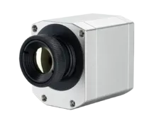
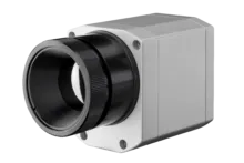
The device integrates an infrared camera, the Optris Xi 400, which plays a crucial role in capturing high-resolution thermal images of the skin. The Xi 400 is equipped with microscope optics, allowing for close-up imaging to detect minute details in skin lesions. It features an uncooled microbolometer detector with a thermal sensitivity of less than 80 mK, making it highly responsive to small temperature changes. The camera captures 16-bit RAW thermal images at a resolution of 382 × 288 pixels and a frame rate of 27 Hz, ensuring detailed thermal data is captured in real time.
The Xi 400’s manual motor focus and the ability to focus at distances between 90 mm and 110 mm enable precise imaging, which is crucial for accurately assessing the boundaries and depths of skin lesions. The device’s design includes a distance holder that maintains a consistent 100 mm distance from the skin, ensuring uniformity in measurements. This precise imaging capability, coupled with the device’s cooling system, allows the HypIRskin to effectively detect and differentiate between malignant and benign skin lesions.
Infrared Thermography in Identifying Melanoma and Carcinomas with the Optris Xi 400 Microscope
Infrared thermography is especially effective in identifying malignant melanoma, basal cell carcinoma, and squamous cell carcinoma—the most common types of skin cancer. When used alongside other diagnostic methods, such as dermoscopy and biopsy, it enhances the accuracy of skin cancer detection and monitoring, leading to improved treatment outcomes.
The successful application of the screening device in clinical research settings highlights its potential as a powerful tool for skin cancer characterization. The device offers rapid measurements, with a total time of approximately three minutes, making it highly practical for clinical use. Its adjustable cooling load and duration enable flexible, case-specific measurements. However, maintaining a homogeneous cooling load is essential to ensure uniform and reproducible results.
The researchers collaborate with Optris, utilizing the Optris Xi 400 infrared camera because it provides the precision and sensitivity necessary for accurately detecting and characterizing skin cancer lesions via infrared thermography. The Optris Xi 400 is equipped with high-resolution thermal imaging capabilities, which are critical for capturing the subtle temperature differences between healthy and cancerous tissues.
This camera’s uncooled microbolometer detector offers a thermal sensitivity (NETD) of less than 80 mK, allowing it to detect very slight temperature variations, a key requirement for identifying the thermal signatures of skin cancer. Additionally, the Xi 400 features microscope optics and manual motor focus, enabling detailed close-up imaging of skin lesions.
Recommended Products


Other Pharmaceutical & Medical Applications


Talk to us about your IR Temperature Measurement Requirements
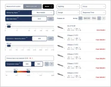
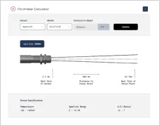
There are over 300 different pyrometer variants to choose from in the Optris infrared pyrometer portfolio each optimized for material, spot size, distance from the target, and environmental conditions. Fortunately, there is a trained engineer to phone or chat with to guide you through the process of choosing the perfect infrared sensor for your application.
The same support is available for the extensive IR camera product line.